All posts by CPTFeditor

Everything you ever wanted to know about radiation and cancer, but were afraid to ask

Julie Bromberg and Laura Covarrubias

Click link to jump to section:

Radiation from the Environment
Medical Devices that Use Radiation
Other Manufactured Devices that Use Radiation
Table: Approximate Dose and Cancer Risk of Various Radiation Sources

When most people think of radiation, they think of manufactured devices such as the nuclear bomb or cancer treatments, which emit high doses of radiation.  In reality, however, radiation takes many forms and is always around us.  Some types are much more dangerous than others.1,2

Most researchers agree that there is no such thing as a dose of ionizing radiation that is so low that it will not have some effect on our body, such as damaging cells.  Usually, the damage is small enough that one dose does not lead to any health problems.  It is likely that the increased risk of cancer from low doses of radiation is so low that studies in the general population can’t detect it.3,4

It is important to note that each exposure to radiation builds up in our body and the risk of cancer increases with each radiation exposure.  So even though a single source of exposure to radiation is unlikely to cause cancer by itself, the combined exposures add up throughout our lifetime and increase our risk of cancer over time.5 This is why it is important to limit unnecessary exposures to radiation.  Radiation exposure during certain sensitive times of development, such as during childhood and puberty, also has more health risks than the same exposures in adults.6,7

Since radiation is always around us, we cannot avoid all radiation, but we can try to limit our exposures.  This article will explain the various risks associated with different sources of radiation and how you can avoid getting too much exposure to radiation.

Radiation from the Environment

Background radiation

Background radiation refers to radiation that naturally occurs in our environment and does not come from any manufactured devices.  Radiation is emitted from the earth, sun, our galaxy, and other galaxies.  Even the human body naturally contains some radioactive elements.8  People who receive few or no high-dose radiation medical tests usually get more exposure to radiation from the natural environment than from any manufactured device.9

This is because we are constantly exposed to a very low dose radiation for our whole lives, while devices such as x-ray tests expose you to radiation for a very short period of time.  On the other hand, one CT scan can be equal to several years of background radiation exposures, so many people receive much higher doses of radiation from medical devices than they do from the natural environment.

The risk of developing cancer from a lifetime exposure of background radiation is about 1 in 100, or 1% of the population.10It is impossible to avoid all background radiation, but the best ways to limit unnecessary exposure to radiation from the environment is to prevent your exposure to radon and repeated unprotected sun exposure.

Radon

Radon is a color
less, odorless gas that comes from decaying rocks and soil.  For the average person, radon accounts for over half their annual exposure to radiation.  Radon comes up from the ground and gets trapped in houses and buildings.

Exposure to a small amount of radon inside is normal, but high levels can cause lung cancer.  Radon is the second leading cause of lung cancer (smoking is the leading cause), and approximately 1 in every 15 houses have too much radon.  The only way to know if you have a safe level of radon in your house is to get it tested.11

Cosmic and terrestrial radiation

Cosmic and terrestrial radiation is radiation that comes from the galaxy and from the earth.  It makes up about 8% of our average yearly exposure to radiation.12 Cosmic radiation includes ultraviolet (UV) rays from the sun that cause tans and sunburns.  UV rays can also damage the DNA in our skin cells and lead to skin cancer.13 Although we cannot avoid UV rays all the time, limiting exposure to direct sun light can reduce your risk of skin cancer.  Tanning beds are also a common source of UV radiation and are just as dangerous as radiation from the sun.  For more information on tanning beds, please read: Tanning Beds: Safe Alternative to Sun?

Being at higher altitudes, such as flying on a plane or living in a “mile high” location, will exposure you to higher levels of cosmic radiation than being at sea level.  While there is technically no “safe” dose of ionizing radiation, the chances of getting cancer from frequent plane trips is very slim.  Studies of airline crew members have not found a significant increase in risk of cancer after many years of working on airplane.14

In addition, living in Denver or other high altitude locations that receive higher doses of cosmic radiation has not been shown to increase the risk of cancer.15

How to Reduce Radiation Exposure from the Environment

Radon:  Test the level of radon inside your house to make sure it is not too high.  You can hire a professional to do this or you can purchase a “do-it-yourself” radon testing kit.  It generally takes only a few minutes and is easy to do.  If there is too high a level of radon in your house, people usually install an active soil depressurization (ASD) system, which is basically a ventilation system.2

UV Radiation: Some of the best ways to reduce your exposure to harmful UV rays are to:

  1. Wear sunscreen (at least SPF 15) year-round in all areas of your body that are exposed to the sun. (However, it is good to get vitamin D from the sun for 15 minutes each day.)
  2. Stay in the shade, especially when the sun is at it’s strongest (between 10am and 4pm)
  3. Wear protective clothing, such as broad-brimmed hats and tightly-woven clothes that cover your hands and legs.

Medical Devices that Use Radiation

X-rays

X-rays use ionizing radiation and are used for many types of diagnostic tests such as CT scans, mammograms, fluoroscopy, and simple x-rays.  These tests allow your doctor to see potential problems inside your body and choose an appropriate treatment.  They can help doctors make life-saving decisions, but some doctors are performing unnecessary scans or are using doses of radiation that are too high.16  Since x-rays use ionizing radiation, they can cause damage to our cells and DNA. X-ray tests can lead to cancer, but several common tests (such as mammograms and bone x-rays) use very low doses that have not been shown to cause a significant increased risk of cancer when administered properly.

Over the past few decades, the average level of radiation that Americans are exposed to has increased rapidly due to increased use of medical diagnostic tests such as x-rays (including dental x-rays and mammograms) and CT scans and cancer treatments.  Diagnostic tests and treatments can help improve patients’ quality and length of life, but there are also risks.  Usually, the benefit of receiving one of these tests outweighs the risk, but patients and doctors need to be wary of performing unnecessary tests, particularly if the test uses high doses of radiation.

Not all imaging tests use radiation that has been linked to cancer. Magnetic Resonance Imaging (MRIs) and ultrasounds do not use x-rays.  Instead, they use non-ionizing radiation and have not been found to increase the risk of cancer or other health problems.17

MRIs and Ultrasounds are a safer alternative to diagnostics tests that use x-rays or other ionizing radiation.

Children, young adults, and fetuses of pregnant women should be particularly careful about getting any x-ray tests.  Children, young adults, and fetuses are more sensitive to radiation, and their young age also allows a longer period of time for cancer to develop.18,19,20

Pregnant women should avoid any x-ray exposure, particularly when they are less than 20 weeks pregnant, since radiation exposure in the womb can lead to mental retardation, growth retardation, leukemia, and other cancers later in life.21  If it is necessary for a pregnant women to be x-rayed, the American College of Obstetricians and Gynecologists states a single x-ray test does not harm the fetus, but a protective lead apron should be used to cover the abdomen.22  However, high-dose, multiple-dose, or x-rays of the pelvic region should be avoided for pregnant women, whenever possible.

Many people get simple x-ray tests, such as an arm, leg, chest, or dental x-ray that look for broken bones or other problems.  Simple x-ray tests use very low doses of radiation,23  and studies have not found an increase risk of cancer among humans who have received a very low dose of radiation.24 Although the dose of radiation used to x-ray different parts of the body will vary, most simple x-rays use less radiation than other types of x-ray scans (such as a mammogram or CT scan).

Mammograms

Mammograms are an x-ray test that is used to detect breast cancer.  This test uses a higher dose of radiation than a simple x-ray, but less than a CT scan or Fluoroscopy.  According to the U.S. Preventative Services Task Force, a woman above age 50 who is at average risk of breast cancer should get a mammogram test once every two years until age 74.25 These new guidelines would expose women to less than half the amount of radiation from mammograms than following previous recommendations (which included annual mammograms starting at age 40 for women at average risk).

While these doses of radiation could cause new cases of breast cancer, the appropriate use of mammograms has resulted in lives saved, and the benefits of getting regular mammograms are likely to be even greater than the risks when the frequency of mammograms is reduced to every other year.

Women who are carriers of the BRCA genetic mutation were previously recommended to begin yearly mammograms at age 25-30, since this mutation puts them at much higher risk of getting breast cancer.  Newer studies have found that starting yearly mammograms before age 35 has no benefit to these women and may instead be harmful.  They end up with higher exposure to radiation over their lifetime, which increases the chance of getting radiation-induced breast cancer that they may not have gotten otherwise.26

Fluoroscopy

Fluoroscopyis an x-ray test that allows doctors to see a continuous x-ray image of your body (like a movie, rather than just a picture as with other x-ray tests).  Fluoroscopy uses an x-ray absorbing dye that is either drunk or injected into the body, which allows doctors to see a better outline of the organ.  This procedure is used to view the digestive system (such as stomach, kidneys, or colon), arteries, or joints.27

Since this test sends x-ray beams over an extended period of time (usually 20-60 minutes),28 it exposes people to much higher doses of radiation than a simple x-ray test, although the doses vary widely depending on the test.

Fluoroscopy and CT scans both use high doses of radiation and pose the greatest and most avoidable risk of radiation-induced cancer.  Limiting the number of CT and fluoroscopy tests you receive is one of the best ways you can avoid getting cancer from radiation.  In addition to increasing cancer risk, this test can damage the skin and cause burns.29

Computed Tomographic (CT) scans

CT Scans are a relatively new type of diagnostic imaging technology that allows doctors to view 3-dimentional pictures of various organs in your body.  CT scans use higher doses of radiation than most other types of diagnostic test and are likely to cause new cancers in some patients, compared to their risk if they had not received a CT scan.

Today, an American’s average lifetime dose of radiation from diagnostic procedures is six times higher than it was in the 1980s.30  This is largely due to the increased use of CT scans. Everyday, 19,500 CT scans are performed in the U.S. and this number continues to climb. Each CT scan is equivalent to 30 – 442 chest x-rays, depending on the dose used for the CT scan.31 One study projected that CT scans performed in the U.S. in 2007 alone will result in 29,000 new cancer cases and roughly 15,000 deaths that would not have occurred if they had not received a CT scan.32 These risks would increase with each additional CT scan a person receives.  Low-dose CT scans, which expose patients to less radiation, are now being used to screen for lung cancer, with concerns about whether the benefits outweigh the risks.  For more information on low-dose CTs, click here.

Unfortunately, there is no established guideline for how much radiation should be used for each procedure.  Different scans require different levels of radiation in order to get a clear image, but some doctors are using more radiation than is necessary.  One study found that different medical facilities had huge variations in the dose of radiation used for the same procedure.  On average, the highest dose given for a CT scan was 13 times higher than the lowest does given for the same type of scan.33 The researchers found no pattern in why this dose variation occurred, and no scientific justification.

Children’s exposure to radiation from CT scans is particularly worrisome because children have many more years to develop cancer than adults receiving CT scans and are more sensitive to the effects of radiation.  A June 2012 study found that children receiving higher doses of radiation — through multiple CT scans– were more likely to develop brain tumors and leukemia than children who had only one CT scan.34  However, brain tumors and leukemia are very rare conditions and the increased risk due to CT scans was relatively small: for every 10,000 CT scans performed on children under 10 years-old, there will be one additional diagnosis of leukemia and one additional diagnosis of a brain tumor. The researchers concluded that the benefits of children having necessary CT scans outweighed the risks of later developing cancer.

A study published in May 2013 looked at children from infancy to 19 years of age in Australia and compared those who had undergone CT scans to those who had never had any.35  Ten years after getting scanned,  there were 24% more cases of cancer among  the 680,000 children and teenagers who had CT scans than among the 10 million children and teenagers who did not undergo CT scans. Young people who had CT scans during the 12-month period before being diagnosed with cancer were not counted because the decision to scan them may have had to do with cancer symptoms.  Children 4 and under had the highest increased risk in cancer, and risk for all increased with the number of scans. The researchers concluded that for every 1,800 people under 20 who had a CT scan, there was 1 additional case of cancer that would not have occurred without the radiation from the CT scan. The CT scans in this study took place between 1985 and 2005 when radiation doses where generally higher than they are today.

Parents should make sure that CT scans ordered for their children are medically necessary and ask their doctors if lower-radiation alternatives exist. While parents should not stop their children from receiving a necessary CT scan because of radiation concerns, they should think about keeping the number of scans below age 20 to a minimum.

The FDA and investigative journalists have also released warnings about occurrence of extreme accidental overdoses of radiation from CT scans.36,37

 In January 2010, the FDA reported that over 250 patients at 4 facilities had received as much as 8 times the amount of radiation that they were supposed to receive.38 Accidental radiation overdoses can result in skin redness, hair loss, increased risk of various cancers and cataracts in the future, and death.  While extreme overdoses of radiation are rare, these avoidable mistakes have lead many health professionals to call for more standardized and comprehensive methods of overseeing medical radiation.39

(Click here for more information on heart CT scans and cancer risk).

Positron Emission Tomography (PET) scans

PET scans differ from other types of diagnostic imaging in that they allow doctors to see how an organ or system is functioning rather than just seeing the structure.  This test works in a very different way than other tests and does not use x-rays-rather, it uses gamma rays, which usually have a higher level of energy than x-rays.

PET scans work by injecting (or swallowing) small amounts of radioactive material, which then spreads throughout the body. The PET scanner is then used to detect the radiation that is emitting from the radioactive material in your body. Procedures that use radioactive material to diagnose and treat patients is referred to as “nuclear medicine.”40

The dose of radiation from a PET scan is similar to CT, and therefore exposes people to a relatively high dose of radiation in comparison to other types of scans.

Using Radiation to Treat Cancer:

Radiation therapy

Radiation therapy uses high doses of radiation to treat various types of cancers.  Beams of radiation are directed at the cancer to kill off cancerous cells.  This can save lives and prevent recurrence of cancer, but healthy cells that are exposed to radiation may develop into a new cancer.  Fortunately, new cancers caused by radiation therapy are not thought to be very common since radiation technology can precisely irradiate a small part of the body that contains cancer, minimizing the amount of healthy cells that are exposed to radiation.41

Another concern about radiation therapy and diagnostic tests are errors in using this technology. Although relatively uncommon, some patients will accidently receive doses that are too high.  In addition to being at increased risk of developing cancer in the future, the incorrect doses can cause serious wounds to the skin, bone, and other organs, as well as death.42,43

Between 1950 and 2006, the frequency of diagnostic radiation increased 10-fold.44 As health professionals continue to find new uses for medical devices that use radiation, people will be exposed to radiation more often. For example, many radiologists have recently started promoting the use of CT scans to screen for colon cancer (known as a virtual colonoscopy), although the FDA has not approved CT scans for this purpose.45

Although many patients would prefer this non-invasive procedure over the traditional direct examinations, it would expose people to high doses of radiation that is roughly equal to 100 chest x-rays (or 3 years of background radiation.46 This means more people are likely to be diagnosed with cancers that are caused by radiation than they would not have gotten otherwise.

Other Manufactured Devices that Use Radiation

Airport and Building X-ray Scans

Backscatter and millimeter scanners have begun replacing metal detectors and are designed to scan a person to determine what weapons or explosives they may have beneath their clothing. Currently, there are about 250 backscatter and 264 millimeter wave scanners in the United States. The TSA hopes to have 1,800 scanners of either type installed by the end of 2014 – which would mean that nearly every airport in the country will have one.

Backscatter scanners look like two large blue boxes:

(Source: Transportation Security Administration)

People raise their arms and stand sideways between these two boxes when they are scanned.

In contrast, millimeter wave scanners look like circular glass phone booths, and the person being scanned stands with their arms raised while part of the scanner rotates around them:

(Source: Transportation Security Administration, from http://www.tsa.gov/videos/travel-tips-advanced-imaging-technology)

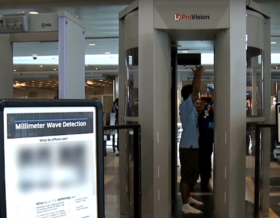
If you are not sure which scanner is in use at your airport, ask a TSA official at the security checkpoint.

While metal detectors and millimeter scans both use non-ionizing radiation, which until recently was assumed to be safe (see our article Can Cell Phones Harm our Health?), backscatter scans use ionizing radiation, which is used in x-rays and known to potentially increase the risk of cancer. Backscatter scans work a little differently from x-rays. X-rays work by sending high-energy radiation to the body and recording the radiation that passes through the body. Dense parts of the body (like bones) block some of the radiation, resulting in lighter areas on the recorded image.  Backscatter scanners also send radiation toward the body, but at much lower energy than an x-ray. Because it is not as strong as the radiation used in x-rays, the radiation does not pass through the body. Instead, the outer layers of the body “scatter” the radiation, which bounces off the body and back toward the machine. People receive most of the radiation that is absorbed by the body is deposited in the outer layers (like the skin and ribs), although a 2012 study showed that radiation from these scans may penetrate to other organs.47  Because the radiation is concentrated in the skin, there are concerns that this could cause skin cancer.

All data on backscatter scans are provided by TSA, a government agency that does not allow independent researchers to examine the machines they use.48 Researchers must therefore make educated guesses using data provided by the TSA, or they must make models of the scanners based on information that the agency releases.

The TSA states that backscatter scans use such low doses of radiation that estimating the potential effects of the scan is extremely difficult.49,50 A 2011 report using information from the TSA found that these backscatter scans expose people to the same amount of radiation that they receive from 3 to 9 minutes of normal daily life or from 1 to 3 minutes of flight.51 To put this into perspective, we would expect only 6 of the 100 million airline passengers each year to develop a cancer in their entire lives due to the backscatter scans.

Dr. David Brenner, a researcher at Columbia University, produced a different estimate based on the risk that the scanners are to the entire population, not just to an individual.52 Dr. Brenner multiplied the risk associated with one scan by the number of scans conducted each year to estimate the number of people who may develop cancer in one year because of the scanners. Because up to one billion scans may be performed each year, Brenner estimated that each year 100 people would develop cancer because of their exposure.

In April 2010, a group of scientists from the University of California, San Francisco wrote a letter of concern to Dr. John Holdren, the Assistant to President Obama for Science and Technology, about the backscatter scans. These researchers pointed out that because backscatter scans only penetrate outer layers of the body, it is possible that these layers receive a higher concentration of radiation than previously believed.

The scientists also expressed concern that sperm may mutate because the testicles are close to the surface of the skin and are exposed to radiation during these backscatter scans. In addition, they noted that the effects of radiation on the cornea (the outer surface of the eye) and the thymus (a part of the immune system located in the chest) have not been studied. While this letter only outlined concerns of the scientists and did not present new data, it called for further testing of backscatter scans.  The scientists called for more rigorous and independent studies to ensure that the scans are safe for the entire population, as well as for all parts of the body.

In a joint reply with the TSA, the FDA stated that the radiation exposures from the backscatter scans were within established legal limits, even for frequent fliers.53 In reply to the scientists’ concerns that the radiation dose to the skin would be higher, the FDA wrote that their calculations showed that a person would have to pass through the scanner 1000 times in a year in order to begin to absorb the annual limit of what is considered safe.54

Not everyone agrees with the FDA, and some people have pointed out that TSA agents operating the scanners may improperly manage the devices or that mechanical errors may occur, either of which could cause the machines to emit more radiation than they are supposed to. From May 2010 to May 2011, there were 3,778 calls for mechanical problems on backscatter machines, but only 2% of those machines were evaluated for radiation safety.55

For a more in-depth look at airport security and radiation, read here.

Microwaves, Cell Phones, and Other Manufactured Devices

There is still much debate among scientists about whether non-ionizing (low-energy) radiation such as microwaves can increase your risk of cancer or other health problems. The concern is not about microwave ovens, but rather the long-term exposures to microwaves from other sources, such as communication towers and cell phones.56 Cell phones emit very low doses of microwave radiation, which was long assumed to be safe.

Although generally safer than ionizing radiation, the much longer-term exposure could make these products potentially dangerous. There are studies indicating that long-term exposures to low doses of non-ionizing radiation can damage DNA and may cause cancers and neurological and reproductive harm.57,58

The rapid increase in use of cell phones and other wireless devices intensifies the need for further research on potential health effects of these non-ionizing radiation sources.  Click here to get more information on cell phones and health.

Tobacco, fertilizer, welding rods, smoke detectors, and several other consumer products also contain some radiation, but radiation from these sources is generally very low (approximately 3% of our yearly radiation dose.[10]

APPROXIMATE DOSE AND CANCER RISK OF VARIOUS RADIATION SOURCES: How does it compare to natural background radiation? What is the lifetime risk of cancer death from one exposure to this radiation source?

Type of Radiation (dose in mSv)† Equivalent Period of Natural Background Radiation‡ Estimated Lifetime Risk of dying from cancer that results from a single exposure§
Airport Security x-ray scanner23 (~0.0001mSv) less than one hour Almost (less than 1 in 100,000,000)
7 hour airplane flight9 (~0.03 mSv) a few days Almost 0 (1 in 1,000,000 – 100,000)
Chest x-ray6 (~0.1 mSv) ~ one week Almost 0 (1 in 1,000,000 – 100,000)
Mammogram (~0.4 mSv) a few months (~2 months) 1 in 100,000 to 10,000
CT of chest27 (~7 mSv) a few years (~2.3 years) 1 in 10,000 to 1,000
Fluoroscopy: colon (barium enema)27 (~8 mSv) a few years (~2.7 years) 1 in 10,000 to 1,000
CT of heart (angiography)27 (~16 mSv) a few years (~5.3 years) 1 in 10,000 to 1,000
PET scan, whole body5 (~14 mSv) a few years (~4.6 years) 1 in 10,000 to 1,000
Fluoroscopy: kidneys, ureters and bladder5 (~15mSv) a few years (~5 years) 1 in 10,000 to 1,000
Whole-body CT scan5 (~22.5 mSv) several years (~7.5 years) 1 in 1,000
Nuclear Medicine: Cardiac stress-rest test (thallium)27 (~40.7mSv) many years (~13.6 years) ~2 in 1,000
Transjugular intrahepatic portosystemic shunt placement27 (~70mSv) many years (~23.3 years) 1 in 100 – 1,000
Lifetime risk of cancer death NOT caused by radiation§§ 1 in 5

†Dose is based on a normal effective dose for that type of scan.  Actual doses used for a specified scan vary widely depending on the medical institution, the individual, and other factors.59

‡Natural background radiation is equal to about 3mSv per year.60

§ Risk of developing cancer is based on EPA cancer risk estimates: “…health physicists currently estimate that overall, if each person in a group of 10,000 people exposed to 1 rem [10mSv] of ionizing radiation, in small doses over a life time, we would expect 5 or 6 more people to die of cancer than would otherwise.”[33]

§§ “In this group of 10,000 people, we can expect about 2,000 to die of cancer from all non-radiation causes. The accumulated exposure to 1 rem [10 mSv] of radiation, would increase that number to about 2005 or 2006.”[33]

All articles are reviewed and approved by Dr. Diana Zuckerman and other senior staff.


Related Content:
Children and cell phones: is phone radiation risky for kids?
Airport security and radiation
Can cell phones harm our health?

Fracking and your health

By Anna E. Mazzucco, Ph.D.
2014


Rig_wind_river

Fracking is in the news, but what is it exactly?  Hydraulic fracturing or “fracking” has boomed in the last few years and is now producing most of the natural gas in the U.S.  It is praised as helping the U.S. be more “energy independent” and has lowered the cost of natural gas.  But, the controversy is whether it can harm our health, especially for people who live near the drilling sites, which are most common in Colorado, Texas, North Dakota, and Pennsylvania.

Fracking uses more than 750 chemicals, some of which are known to harm human health.61,62,63   But since fracking is a relatively new technology in the U.S., scientists are still trying to understand how fracking chemicals get into the air and drinking water in nearby areas.

Of the more than 750 chemicals used in fracking, more than 100 can affect our hormones.  These chemicals are called endocrine-disrupting chemicals, and research published in 2011 shows that these chemicals can get into the water near fracking sites, where they could potentially cause infertility, diabetes, and cancer.64 This study in Colorado found higher levels of endocrine disrupting chemicals in water samples from areas with more drilling sites compared to areas with fewer sites.  Many of the water samples were found to contain chemicals that either mimicked or blocked estrogen (a hormone that is high in females) or androgen (a hormone that is high in males).   In fact, 89% of the samples taken near fracking sites had chemicals which increased estrogen, 41% had chemicals that blocked estrogen, 12% had chemicals that increased androgen, and 46% had chemicals that blocked androgens.  In contrast, water from areas far from fracking rarely had chemicals that affect hormones.  The researchers also found small increases in these chemicals in the Colorado River, probably from the fracking sites.  This could spread the chemicals into a larger area.

What kind of impact could these chemicals have on human health?  When children are exposed to high levels of hormones, this could cause early puberty in children, or abnormal sexual development in a fetus.  Chemicals that block hormones could also affect sexual development or fertility.  In adults, this exposure could increase the risk of infertility, obesity, diabetes, or certain types of cancer, such as breast cancer, testicular cancer, and vaginal cancer.   Serious diseases, and especially cancer, can take years to develop, so it is important to study families in fracking areas carefully for many years to see what happens.  Of course, meanwhile the families could be harmed if they are not protected from these chemicals in their water.

Two other reports have raised concern about fracking and the health of newborn babies.  These reports provide evidence that mothers living near fracking sites are more likely to give birth to newborns who are underweight.  One study found that babies were 25% more likely to have low birth weight if they were born to mothers living near fracking sites compared to babies born of mothers who didn’t live near one.65 A second study found that the chance of a low birth weight baby was more than 50% higher for babies born near fracking sites, compared to babies born to the same mothers in a location far from fracking.66   This study was presented at the annual meeting of the American Economic Association in January 2014.  Neither of these two studies has been published yet.

To be more conclusive about the health risks of living near fracking sites, we need large public health studies that include information from medical records and residence history.  Currently, most of the information we have is based on evidence of surface and ground water contamination from fracking sites.67,68

Meanwhile, several families and communities have already sued because of their concerns about health problems related to fracking, and the Environmental Protection Agency (EPA) and state agencies have investigated drinking water contamination in West Virginia, Pennsylvania, Wyoming and Texas.  The EPA is currently working on a report on the potential impact of fracking on the U.S. water supply, with a draft expected late in 2014.  Legislation to lower health risks due to fracking has been proposed in many states, including California and Illinois.  In 2012, Vermont became the first and only state to ban fracking.


The worst new drug of 2014

DIANA ZUCKERMAN, PHD, PRESIDENT OF The Cancer Prevention & Treatment Fund 

It’s only February, so it may seem early to be talking about one of the Food and Drug Administration’s (FDA’s) worst decisions of 2014. Yes, the year just started, but the FDA has already made a decision that could potentially harm thousands of patients.

The agency just approved a new diabetes medication that doesn’t noticeably improve health but may in fact cause cancer.

It’s called Farxiga (Dapagliflozin) and it will be available—and probably widely advertised—very soon. The good news is that the drug lowers blood sugar, which is a major symptom of type 2 diabetes. However, lowering blood sugar is not necessarily a way to improve health. The new drug is meant to reduce medical problems such as heart disease or kidney damage. Unfortunately, there is no evidence that it actually improves health—in fact, quite the opposite.

The FDA reports that the most common side effects of Farxiga are genital fungal infections and urinary tract infections. Not fun, but those aren’t its most serious side effects. If a patient has moderate or severe kidney damage (as many diabetics do), the drug is not beneficial and could cause further damage, possibly even renal failure.

But the more frightening news is that patients taking Farxiga in studies done for the FDA were more than five times more likely to contract bladder cancer than the patients who took an older diabetes drug. Based on the findings of the companies making and selling Farxiga, FDA scientists concluded that the drug might stimulate bladder cancer in patients already at risk. The fear is that patients take diabetes drugs for years, and the longer someone takes this drug, the greater their risk of cancer.

Bladder cancer can be fatal, and is especially dangerous in people who have diabetes.

The companies’ studies also found that patients taking the drug were more than twice as likely to get breast cancer as diabetics not taking the drug, but they say this could have happened by chance.

It’s important to mention that only a small number of African Americans were studied. Because African Americans are more likely to have diabetes than Caucasians, these studies should have included greater numbers of them to find out if the drug is safe or effective for them.

With so many other diabetes drugs already on the market, why would anyone want to take this drug, and why would FDA approve it?  Here’s the scorecard, based on information provided by FDA scientists and available on the FDA website:

  1. Is the drug new? Yes.
  2. Does it reduce the medical problems caused by type 2 diabetes, such as blindness, heart disease, kidney damage, or amputations? No.
  3. Can it cause kidney damage or make it worse? Yes.
  4. Does research show that patients taking it are more likely to get bladder cancer. Probably.
  5. Does research show that patients taking it are more likely to develop breast cancer? Maybe.
  6. Does research show it is more effective than most diabetes drugs on the market? No.
  7. Will it cost more than most other diabetes drugs on the market? No information on cost yet, but new drugs usually do cost more.

The FDA is requiring that the label for the drug warn patients with bladder cancer that they shouldn’t take Farxiga because it might make their cancer worse. Ya think? And, the agency is requiring the companies to study 17,000 diabetes patients for at least four years to determine whether and how often patients taking Farxiga are diagnosed with cancer, liver problems, or heart disease when the drug is taken for a longer period of time.

The FDA rejected this drug two years ago because of these cancer concerns and questions about how well the drug prevents the major risks of diabetes. But the FDA has been beaten up by many complaining congressmen since then, and by some patient groups, pushing it to approve more new drugs quickly rather than waiting for safety studies to be completed. Also, the pharmaceutical companies don’t like to take no for an answer, they spend lots of money on lobbying, and they are very effective at pushing FDA to reconsider rejections. Because of that, the FDA selected an advisory committee to review the data in December, and those doctors and scientists also expressed concerns about risks and benefits. I was at the meeting and pointed out that there were too many serious unanswered questions.

Despite my concerns, the concerns of FDA scientists and other experts, and the concerns of the advisory committee members themselves, the committee recommended that if the companies continued to study the drug after it was approved, in order to figure out what the actual risks are, the company could sell it now.

It’s too early to say whether this will be the worst decision the FDA makes in 2014. Maybe the diabetes patients who take it will be lucky and not be harmed by the drug. Or maybe only a few patients will develop cancer or kidney failure as a result of taking it. Or the FDA scientists who expressed their concerns could be wrong, and I could have made a mistake when I agreed with them. But regardless of what happens with this drug, it still would be a bad decision to allow the sale of a drug with such serious (potentially fatal) unanswered questions.

Given the epidemic of diabetes in this country, the risks are huge. For that reason, this drug worries me even more than some of FDA’s similarly mind-boggling decisions of 2013, such as:

  • drug for hot flashes that has no meaningful benefits but can increase the risk of suicide among women who weren’t depressed
  • An antibiotic for pneumonia that seems to kill more patients than other antibiotics

I’ve always respected the scientists at the FDA for working so hard to try to keep all of us safe. I still do. But decisions are being made at the agency that seem to ignore scientists and science, and that is dangerous for all of us. Whether we have diabetes, hot flashes, pneumonia, cancer, or any other disease, we deserve better.


A version of this post appeared in The Huffington Post, Maria’s Farm Country Kitchen

 In 2018, the U.S. Food and Drug Administration (FDA) announced a new warning on Farxiga and other SGLT2 inhibitors because they can cause a rare but life-threatening infection of the genitals and area around the genitals. The infection is called necrotizing fasciitis of the perineum or Fournier’s gangrene.  

Fast Facts: Should you get screened for Lung Cancer?

For our in-depth article on lung cancer screening, click here.

By Tiffanie L. Hammond, Amrita Ford, MA, and Anna E. Mazzucco, Ph.D.
January 2014

Lung cancer is the second most commonly diagnosed cancer in both men and women in the U.S., but it is the #1 cancer killer. In 2013, approximately 228,000 men and women were diagnosed with lung cancer and close to 160,000 men and women died from it.  One of the reasons lung cancer is so deadly is that symptoms usually appear during the later stages, when treatment is least effective. General symptoms include:

  • a persistent cough that may worsen over time, including coughing up blood
  • breathing trouble, such as shortness of breath
  • chest pain
  • raspy or hoarse voice
  • frequent lung infections, such as pneumonia
  • extreme and constant fatigue
  • unintentional weight loss

If you experience any of these symptoms, call your doctor.

The purpose of screening for cancer is to diagnose it before symptoms appear, when it can be treated more effectively.  In July 2013, the U.S. Preventative Task Force (USPTF) released a draft recommendation for screening those at highest risk for lung cancer using low-dose computerized tomography scans (low-dose CT), which was finalized in December.  CT scans provide very detailed pictures of your lungs, much more detailed than x-rays. Researchers found that using low-dose CT scans could reduce lung cancer deaths by 20%.  People who should be screened include:

  • Current smokers between 55 and 79 years-old who have a smoking history of “30 pack-years” (20 cigarettes a day for 30 years, 40 cigarettes a day for 15 years, and so on)
  • Former smokers (who quit in the last 15 years) who are between 55 and 79 years-old and had a “30 pack-year history of smoking.


To calculate your pack years, visit http://smokingpackyears.com/.

This draft recommendation was finalized at the end of 2013. Already insurance companies are covering the cost of the screening, as long as the person meets the screening criteria.

As with any screening, there are risks as well as benefits. Some of the risks include radiation exposure and a high rate of false positive test results—test results indicating that a person has something worrisome in their lung that turns out to be nothing serious. Some experts call this “over-diagnosis.” In the largest study done so far, about 1 in 4 people had a false positive finding if they had three years of annual screening.  A false positive finding can lead to invasive procedures, such as needle biopsies, which can puncture the lung and cause it to collapse.  Although very serious complications are rare (less than one in 1,000), they can occur.  Even low-dose CT scans of the lungs expose patients to much more radiation than a chest x-ray. There is almost no short-term risk from this radiation, but having many x-rays or several CT scans can increase a person’s risk of cancer. A 2012 law increased funding for lung cancer research to promote better detection and treatments in the future.   Research is also being done to find ways to reduce the number of false-positives and unnecessary follow-up procedures.

For more detailed information about lung cancer risk factors, symptoms, detection, and CT screening, including the risks of radiation and over-diagnosis, see this article.

Clinical evidence in FDA drug approvals varies widely, study finds

by Sabriya Rice, Modern Healthcare
January 21, 2014

Not every new drug approved by the U.S. Food and Drug Administration has undergone the rigorous clinical testing that physicians and their patients might expect, according to new research.

A study published Tuesday in the Journal of the American Medical Association finds that the FDA has “flexible standards” for approving of new therapies. Using publicly available information from the FDA drug database, investigators identified 188 novel therapies for treating 206 conditions approved between 2005 and 2012. Of these, 37% were approved on the basis of a single clinical trial, 38% on the basis of two trials and 25% had been tested in three or more trials.

Although most therapies were supported by at least one randomized, double-blind trial—the gold standard for clinical research—there was wide variation in the duration, size and completion rates. Comparative-effectiveness data was available for less than half of the indications.The purpose of the study, according to its authors, was not to suggest that the FDA is not rigorous in its approach to drug reviews. The regulatory flexibility allows for a customized approach, and the agency can rapidly approve potentially effective therapies for life-threatening diseases and those for which there is no existing, effective treatment, such as orphan diseases, said Dr. Joseph Ross, assistant professor of general internal medicine at Yale University School of Medicine and one of the study authors.

According to Ross, the drug approval variability is problematic in the sense that both patients and physicians feel the research is the same across all drugs approved.

The FDA, in a statement responding to the findings, explained that drugs may be tested in clinical trials that enroll hundreds of participants, while others, particularly those seeking to treat rare diseases, may be tested in trials that enroll only a handful of participants. “In all cases, however, the statutory standards of safety and efficacy must be met in order for the drugs to be marketed in the United States,” the agency said.

Whether or not the process has become too lenient has become a topic of debate among advocates.

The Progressive Policy Institute, a center-left think tank, acknowledged in a policy brief (PDF) that the FDA must strike a difficult balance. “If it is too lenient, (the FDA) will allow the sale of drugs and medical technology that could harm vulnerable Americans. Too tight, and the U.S. is being deprived of key innovations that could cut costs, increase health, and create jobs.”

Some argue, however, that physicians don’t have time to sift through statistical data on every new drug approved, and when they do, there is little information to choose from as they make decisions about the safety of new therapies.

“With new drugs, there is often an exaggeration of the benefits and underreporting of the risks,” said Diana Zuckerman, president of the National Research Center for Women & Families, who has testified at several hearings on drug safety. “There’s so much emphasis on drugs being the latest, the most innovative and novel—but unfortunately this usually means it’s just new, not necessarily better,” she said.

Zuckerman conceded that it’s a given that the FDA should have the flexibility to provide access to new treatments when there are no available options. “But the FDA shouldn’t be rushing studies for diseases that have good alternatives. It’s better to have an old treatment that is proven to be safe and effective than a new treatment that we don’t know is safe and may not improve health.”

“I think we can all agree that if you have a disease for which there are no available treatment options, that is the time to be flexible, although you’d still want the best possible research,” she said.

Ross, the Yale researcher who worked on the study, encourages physicians to be more nuanced with how new treatments are presented to patients. When prescribing newly approved drugs that have limited trial data, he said physicians should be clearer with patients about what the research shows. The physician should say, “There are not a lot of options, this drug was just approved, but we don’t know if it extends your life,” rather than giving the patient the impression the drug does things for which it has not been tested.

To view the original article in Modern Healthcare, click here.

Comments of the Cancer Prevention and Treatment Fund on FDA draft guidance to industry on Acrylamide in foods

January 14, 2014

The Cancer Prevention and Treatment Fund strongly supports the Food and Drug Administration in its efforts to advise industry on reduction of acrylamide in food products.  The Grocery Manufacturers Association estimates that acrylamide is present in approximately 40% of the total caloric intake in a typical American diet.69 Given this near-ubiquity, and the fact that the chemical reaction which produces acrylamide also produces commercially desirable color, taste and texture characteristics, reduction of acrylamide represents a challenge.  However, the evidence of possible human harm necessitates its treatment as a significant public health issue. While this report represents an important step in FDA regulation of acrylamide and suggests many possible acrylamide reduction methods, we are concerned that this guidance is not specific enough in providing clear and concrete recommendations that can be implemented.  Although FDA guidance does not have the force of law or regulation, the addition of terms such as “when feasible” implies that the FDA is not serious in its efforts to persuade companies to substantially reduce acrylamide.  FDA monitoring of acrylamide in 2002 indicated wide variation even among products from the same food category –as much as 5 or 10 fold differences in several categories.  This is clear evidence that significant acrylamide reduction can be accomplished without losing desirable product qualities. Thus, although there is clearly much room for improvement, this report contains few immediately implementable guidelines for industry. Since 2002, it has been known that acrylamide is created in food products as the result of a reaction between carbohydrates and the amino acid asparagine at high temperatures during browning (i.e., the Maillard reaction).70  In addition to its known neurotoxic properties, both animal toxicology and human epidemiological studies suggest that acrylamide may be cancer-promoting, which has led to its carcinogen classifications by EPA, NTP and IARC.71,72,73,74 Higher dietary acrylamide consumption has been associated with increased risk of endometrial, ovarian, pancreatic, renal and possibly breast cancer.75,76,77,78.  Acrylamide is already regulated in drinking water and was classified by EPA as “likely to be a carcinogen to humans” and was classified by the National Toxicology Program as “reasonably anticipated to be a human carcinogen,” both more than a decade ago.  The European Food Safety Authority has been overseeing acrylamide monitoring within the European Union since 2007, and the European Commission has set recommended indicative values for acrylamide in food products, providing a quantitative framework for both assessment of reduction efforts and investigative action.  This is a very important health issue and we strongly urge the FDA to intensify its efforts and assert leadership of both the national and international efforts to regulate acrylamide and ensure public safety. Our areas of specific concern are the following:

  • While encouraging manufacturers to conduct their own testing, FDA should update and expand its own monitoring efforts.  Monitoring of acrylamide in food products over time is needed for any reduction efforts to be assessed and successfully implemented.  The current FDA monitoring strategy tested only several hundred foods in four geographic regions annually between 2002 and 2006, and the last publicly available information is from 2006.79  Given the wide range of acrylamide levels even within a single food category, more extensive and up-to-date monitoring is needed to adequately evaluate acrylamide levels and the success of reduction methods.
  • While this report encourages manufacturers to monitor acrylamide levels, it does not give any specific values which should prompt corrective efforts.  Without such guideposts, monitoring alone is unlikely to result in significant reductions.  Recommended target values or action levels, together with active monitoring, will allow FDA and manufacturers to directly access efficacy of reduction efforts and trigger investigation when needed.  The European Commission has set indicative values for acrylamide in food products, including separate values for products intended for infants and young children, and these values are intended to be gradually reduced.80  Indeed, these indicative values have been broadened to include more specific categories and some have already been lowered since their release in 2011, and the European Food Safety Authority is currently conducting a risk assessment at the request of the European Commission to determine if current recommendations are sufficiently protective.  Such a system provides a quantitative framework for reduction efforts and allows increased surveillance of items of special health importance.  Values at least as low as the 2013 European Commission indicative values should be adopted, with the shared intent of gradual lowering of these values as reduction efforts improve.
  • Without accurate and affordable detection techniques, manufacturers are unlikely to measure acrylamide in their products, especially when participation is voluntary.  This guidance encourages manufacturers to be aware of acrylamide levels in their food products.  This is a crucial step towards evaluating reduction efforts.  However, this vital imperative is followed by a discussion of both the technical limitations and expense associated with current methods of acrylamide detection.  While FDA has committed to improving these techniques, they remain costly and fraught with technical limitations, making widespread voluntary use, especially by small manufacturers, unlikely.  FDA should continue to investigate means to improve acrylamide detection and make specific recommendations to industry regarding best possible techniques in order to facilitate participation in monitoring.  As an example, the European Commission has recently set measurement uncertainty (MU) values and tasked the European Committee for Standardisation (CEN) with analytical standardization of LC-MS and GC-MS for acrylamide detection.  Such efforts, in addition to adoption of standard references, will increase consistency and improve confidence in acrylamide detection efforts.
  • In this guidance, the FDA specifically states that it does not intend to recommend one method over another.  This is unfortunate because it leaves both guesswork and legwork to industry.  FDA states that “this guidance is intended to suggest a range of possible approaches to acrylamide reduction and not to identify specific recommended approaches.”  The role of federal agencies should include evaluating reduction approaches to determine which are more efficacious and feasible than others, and providing that potentially useful information to industry, even if only to help identify and encourage prioritization of those approaches first, in addition to continuing research in this area.  Clear communication of superior and cost-effective approaches to acrylamide reduction may result in higher industry participation and more successful reduction efforts.
  • FDA monitoring since 2002 has shown that many foods contain higher levels of acrylamide than the level considered safe by the EPA for drinking water.  Some of the highest acrylamide levels are found in potato and cereal products which are common in the American diet.  These surveys also show that a healthy diet which includes whole grains can have significant acrylamide levels, potentially even higher than a diet which includes less healthful choices such as potato chips and French fries.  The FDA has maintained its message to consumers that a balanced, healthy diet is a way to manage concern over acrylamide consumption, when the evidence shows that this advice is not accurate. 
  • The effects of acrylamide reduction on overall product nutrition should be considered in the context of all health risks and benefits.  For example, lower temperature frying reduces acrylamide, but also requires longer cooking time, resulting in higher fat content in fried foods.  While we commend thorough consideration of all possible health implications of acrylamide reduction methods, FDA should also consider which outcomes can be more easily mitigated by other dietary or lifestyle interventions in order to fully assess risks and benefits.

Lastly, as acrylamide accumulates in food as a result of the handling and cooking process, rather than in the raw food itself, and is a serious human health concern, it could be viewed as source of food adulteration and regulated as such under Section 402(a) of the Federal Food, Drug and Cosmetic Act with action levels.  We ask that FDA consider these improvements to this draft guidance, and use its full authority to ensure that the public is sufficiently protected.

The Cancer Prevention and Treatment Fund

 For additional information, contact Anna Mazzucco at am@center4research.org or (202) 223-4000.

Do high blood pressure and extra weight make prostate cancer deadly?

By Jessica Cote, BS and Anna Mazzucco, PhD
2014

Prostate cancer is the most common cancer and the second leading cause of cancer-related deaths in men in the United States. One in six men will be diagnosed with prostate cancer in his lifetime, with about 90% of cases occurring in men 55 and older, and 71% of deaths occurring in men 75 and older.81

Even though prostate cancer is a leading cause of death, most prostate cancers are not very dangerous. Two key facts are important to remember:

  1. Many older men who aren’t diagnosed with prostate cancer have the disease but will never be harmed by it.
  2. Because prostate cancer usually grows very slowly, most men who are diagnosed with prostate cancer will die of something other than prostate cancer. A 2012 study showed that only 16% of men in the U.S. diagnosed with prostate cancer died from this disease.82

Although the death rate is relatively low, it is important to find ways to prevent prostate cancer deaths with treatments that do not have serious side effects.

The cause of prostate cancer is unknown, but older men, African-American men, men who drink high amounts of alcohol, farmers, and men who were exposed to Agent Orange pesticides are all at higher risk.83  The high incidence of prostate cancer in Western Europe and North America is thought to be related to a “Western” diet, which is high in refined grains, other processed foods, and saturated fats.84  Meat and dairy products tend to have more saturated fats than other foods, and red meat has more saturated fat than chicken or fish.

Does weight increase the risk?

A 2014 report indicates that being obese can increase a man’s risk of advanced prostate cancer.85  The report was based on meta-analyses that combined and analyzed the results of several large studies. The report analyzed the impact of obesity that was measured three ways, by body mass index (BMI), waist circumference, or waist-hip ratio (WHR), and found a similar link.  This suggests that preventing potentially fatal forms of prostate cancer is another reason to maintain a healthy weight.

What is metabolic syndrome and does it increase the risk of dying from prostate cancer?

Metabolic syndrome refers to a group of factors that increase the risk of coronary artery disease, stroke, and type 2 diabetes.86 The syndrome includes insulin resistance, obesity (especially extra weight around the middle and upper body parts), high blood pressure, and high levels of blood sugar and fats.

Recent research tells us that metabolic syndrome is also related to prostate cancer deaths. A 2012 study of the medical records of more than 289,000 men, published in 2012 in the journal Cancer, found that the risk of getting prostate cancer didn’t seem to be affected by metabolic factors, but the risk of dying from prostate cancer was.87  During a 12-year period, 6,673 men were diagnosed with prostate cancer and 961 died from the disease. Men with a higher BMI, elevated blood pressure, and a high composite metabolic score (from BMI, blood pressure, and blood levels of glucose, cholesterol and triglycerides) were more likely to die from prostate cancer than other men.

The study was well-designed with a large sample size and health information that was collected from patients during many medical exams. However, like most studies, this study wasn’t perfect. Researchers did not record details about the prostate cancer such as tumor stages and patterns of spreading, nor did they consider family history of cancer, medication, socioeconomic status, or other diseases that may have occurred in addition to prostate cancer. All those factors could have influenced the chances of the men dying of prostate cancer. Even so, with hundreds of thousands of men in the study, it is likely that the results should be taken seriously: men can increase their chances of surviving prostate cancer (as well as heart disease) if they reduce metabolic problems.

When we consider this study as well as the 2014 report together, it provides convincing evidence that obesity increases men’s risk of dying of prostate cancer.   While prostate cancer is common among all men, being obese and having the common problems associated with obesity (such as high blood pressure and cholesterol) may make the cancer more aggressive.

What else should men do to lower their risk of getting and dying from prostate cancer?

Cancer screening seems like the best way to reduce the risk of cancer, but that is only true if the screening test is accurate and the treatment is safe and effective.  In 2012, the U.S. Preventive Services Task Force recommended against prostate cancer PSA screening tests for men of any age if they do not present any symptoms of prostate cancer (see: Are Annual Prostate Cancer Screenings Necessary? Should Early Stage Prostate Cancer Be Treated?). The Task Force was convinced that the benefits of PSA-based screening for prostate cancer did not outweigh the harms.88

Even though screening isn’t helpful for men with no symptoms of prostate cancer, it could be very effective at saving the lives of men with symptoms.  If you have one or more symptoms of prostate cancer (trouble urinating, blood in the urine, discomfort in the pelvic area), talk to your doctor about getting a PSA or other test for prostate cancer.

Bottom line: If you’re a man over 50, even if you’ve never been diagnosed with prostate cancer and aren’t presenting any symptoms, the latest research tells us it’s a good idea to do the following:

  1. Eat a diet low in saturated fats. This means limiting intake of high-fat cheeses and other dairy products and choosing leaner cuts of meat.
  2. Watch your intake of sugars and starches– this includes beer, wine and alcohol of all kinds. While 1-2 drinks a day can lower your risk of heart disease and possibly reduce your risk of stroke, more than that can increase your risk of diabetes or related metabolic problems.89
  3. Weigh yourself regularly.  Most men don’t, but if you weigh yourself frequently, it will help you keep your weight down.  If you gain a few pounds, you should eat less and exercise more until your weight is back down.
  4. If you have type 2 diabetes and have been prescribed a special diet or medicine, be sure to stick to it and take your pills as directed.

Early stage breast cancer: a patient and doctor dialogue

Common Questions About Early Stage Breast Cancer:

What type of tumor do I have? What does “invasive” mean?

What does “lobular” mean? What does “ductal” mean? What does it mean for my treatment?

What is an “early stage” breast cancer?

What’s my chance of surviving this cancer with each treatment?

Why would any woman pick mastectomy if the survival rate is the same (or not quite as good) as lumpectomy?

What is the difference in survival rates between lumpectomy with radiation and mastectomy?

Does the decision about what kind of surgery to have affect whether I need chemotherapy?

I have breast cancer in my family. Should I choose the more aggressive treatment? Should I undergo surgery to prevent breast cancer?

What are the chances of the cancer coming back if I get a lumpectomy with radiation? If it comes back, is it likely to be invasive? If I decide on a lumpectomy/radiation, how can you be sure there are no other “spots” in the breast? Wouldn’t a mastectomy eliminate that possibility?

What does “margin” mean?

I have heard that some tumors are “estrogen receptor-positive?” What does that mean? If my tumor is estrogen receptor-positive, should that make a difference in my treatment?

If I choose a lumpectomy, how much of my breast has to be taken out? Will it affect the look of my breast? What will the scar look like?

What will my breast look like after lumpectomy/radiation? I hear it gets hard.

I thought that radiation can cause cancer. Will it increase my risk for other cancers?

Can I have a mastectomy without removing the nipple?

What are the side effects of both surgical treatments? What’s the worst case scenario?

Can I have breast reconstruction at the same time as my mastectomy?

With reconstruction, can I change the size of my breasts? Can the plastic surgeon make the other breast match?

What happens when each treatment ends? How often do I see you?

If I have a lumpectomy and I get a recurrence, will I have to have a mastectomy then? Can I have reconstruction after radiation?

These are questions that breast cancer patients commonly ask their doctors. What’s your recommendation? What treatment would you recommend if I were your wife/sister/daughter? What do most of your patients in my situation decide?

Should I get a second opinion?

What type of tumor do I have? What does “invasive” mean?

A “tumor” is an abnormal growth that can be “benign” or “malignant.” Benign breast tumors do not threaten life and do not spread to other parts of the body. Malignant breast tumors are cancers that may threaten life and may spread to other parts of the body. A malignant tumor that grows into surrounding tissues is called “invasive.” Invasive tumors are more likely to spread to other parts of the body than non-invasive tumors.

What does “lobular” mean? What does “ductal” mean? What does it mean for my treatment?

Each breast is composed of up to 20 sections called “lobes.” Each lobe is made up of many smaller “lobules,” where milk is made. Lobes and lobules are connected by small tubes called “ducts” that can carry milk to the nipple.

Lobular carcinoma in situ (LCIS) is a benign tumor that consists of abnormal cells in the lining of a lobule. Even though “carcinoma” refers to cancer, LCIS is not a cancer and there is no evidence that the abnormal cells of LCIS will spread like cancer. Instead, having LCIS means that a woman has an increased risk of developing breast cancer in either breast. Despite the increased risk, most women with LCIS will never get breast cancer. No treatment is necessary and surgery should not usually  be recommended for LCIS because the LCIS cells do not become cancer. Occasionally women with LCIS choose bilateral mastectomy as a preventive measure, but most surgeons consider this inappropriate.  Some women undergo lumpectomy, but experts believe that surgery has no benefits except if the LCIS is a particularly dangerous type.  Some women choose to take tamoxifen to decrease the likelihood of breast cancer. LCIS is sometimes called “Stage 0″ breast cancer, but that is not really accurate because it is not really cancer.

Ductal carcinoma in situ (DCIS) is made up of abnormal cells in the lining of a duct.  It is also called intraductal carcinoma. The abnormal cells have not spread beyond the duct and have not invaded the surrounding breast tissue.

Some experts question whether DCIS should be called cancer, because it does not spread and is therefore not dangerous.

Most of the time, DCIS is not harmful.  Experts estimate that if the DCIS is not surgically removed, approximately 1 in 3 DCIS patients will find that their DCIS will change into the kind of cancer that is invasive. A  Consensus Conference held at the National Institutes of Health (NIH) in 2009 concluded that most women with DCIS are eligible for breast-conserving surgery and that very few require mastectomy. Radiation therapy after lumpectomy helps prevent recurrence of DCIS and the development of invasive breast cancer. If the DCIS is spread out or is in more than one location in the breast, some women will choose to undergo a mastectomy. In the treatment of DCIS, underarm lymph nodes usually are not removed with either breast-conserving surgery or mastectomy. Tamoxifen or another hormone therapy is sometimes used in combination with one of these two surgical treatment options.

DCIS is sometimes called Stage 0 breast cancer or “pre-cancer” because it is not invasive.

What is an “early stage” breast cancer?

Invasive breast cancer is categorized as Stage I, II, III, or IV. Stages I and II and IIIA are considered “early stage” invasive breast cancer and generally refer to smaller tumors that have not yet spread to distant parts of the body.  The cancer may have spread to lymph nodes under the arm.

After the health professional explains surgical options, such as breast-conserving surgery (often called lumpectomy) with radiation, modified radical mastectomy, or simple mastectomy, these are the questions most patients will want to ask.

What’s my chance of surviving this cancer with each treatment?

Most women who are newly diagnosed with early-stage breast cancer have a choice: breast-conserving surgery (such as lumpectomy) or a mastectomy (also called a modified radical mastectomy). The decision is not between your breast and your life. Women with early-stage breast cancer who undergo breast-conserving surgery with radiation therapy live just as long as those who undergo mastectomy. In fact, the latest research shows that women who undergo lumpectomy with radiation tend to live longer than women of the same age and type of diagnosis who underwent mastectomy.

When the patient is told that the survival rate for lumpectomy with radiation is the same as for mastectomy, some women may be surprised or skeptical.

Why would any woman pick mastectomy if the survival rate is the same (or not quite as good) as lumpectomy?

Thanks to early detection, between 70 and 75 percent of women diagnosed with breast cancer today are possible candidates for lumpectomy or other breast-conserving surgery. Yet, half of these women undergo mastectomies instead. Some of those women are making a well-informed choice. Some do not know that they have a choice. And, because of the costs of health care, some cannot afford to make the choice they would prefer.

Unfortunately, cost sometimes prevents women from choosing breast-conserving surgery. Lumpectomy followed by radiation costs more in the short-term than mastectomy, and some insurance plans do not cover all the expenses of the lumpectomy or the radiation therapy. Reconstruction of the breast after mastectomy adds to the cost, but the law requires that insurance covers that expense. Despite the slightly higher cost of lumpectomy and radiation, that choice is actually less expensive if you look at costs for the five years after the initial diagnosis. Lumpectomy preserves the breast and there are few additional costs when the radiation treatment is completed, whereas breast reconstruction after a mastectomy may require several surgeries that add to the cost over time. This information may help women who are concerned about cost to decide what is best for them.

Another reason why women choose mastectomies is because they do not want to undergo radiation therapy or are unable to arrange radiation treatments. Radiation therapy is usually an outpatient procedure performed over a period of at least 5 weeks, and some women are not able to make that commitment. Some women live far away from radiation facilities, or can’t afford to take the time for daily treatments. Others may have health conditions such as lupus or heart disease that prevent them from undergoing radiation. Since radiation reduces the chances of recurrence for women choosing lumpectomy, it is important that patients and their doctors consider the required time commitment to radiation therapy before deciding which surgical procedure is best for them.  Fortunately, there are newer types of internal radiation that are more convenient, but unfortunately there is less information about their effectiveness compared to traditional radiation treatment.

Fear is another reason why some women choose mastectomy. Some women are afraid of radiation therapy. Radiation therapy does cause side effects, but they are usually mild—like fatigue or skin irritation. Only very infrequently does radiation therapy induce more severe side effects.

Fear of recurrence of breast cancer is another reason why some women prefer a mastectomy to a lumpectomy. Some women assume that breast cancer won’t return if the breast is removed. However, women may have a recurrence on the chest wall where the breast was removed because some breast tissue remains even following a mastectomy. For women who choose breast-conserving surgery with radiation, research clearly shows that radiation reduces recurrence for most women with early-stage breast cancer. The risk of cancer returning in the same breast is very low. After 12 years, only one out of approximately 10 women will have had a recurrence of cancer in the same breast. Most importantly, even if breast cancer does recur in the same breast, that does not reduce the woman’s chances for a healthy recovery. However, a recurrence could require additional surgery, and a woman may decide to have a mastectomy at that time.

Many women want to make the surgical choice that will enable them to “get it over with and get on with my life.” Many of these women choose mastectomies, in order to avoid the several weeks of radiation that is required for lumpectomy patients. However, even mastectomy patients may find that recovery takes longer than expected. Lymph nodes are removed with both lumpectomy and mastectomy, and the pain from arm swelling that can result may last a long time and be debilitating. If chosen, breast reconstruction after mastectomy often requires multiple additional surgeries and significant recovery time. Breast implant manufacturers have informed the FDA that one in four patients whose breasts were reconstructed with implants have at least one additional surgery within three years. For women undergoing TRAM flaps and other reconstruction procedures, the pain from surgery can last for months.

What is the difference in survival rates between lumpectomy with radiation and mastectomy?

In a study of more than 112,000 women with early-stage breast cancer, the women who had lumpectomies and radiation were significantly more likely to be alive and less likely to have died from cancer or other causes.  This study followed women for an average of 9 years after their diagnosis. “Statistically significant” means that this difference was very unlikely to have occurred by chance.  Of course, the results could be different 20 or 30 years later, but that seems unlikely.

Does the decision about what kind of surgery to have affect whether I need chemotherapy?

Chemotherapy is not recommended for most women with early stage breast cancer. If chemotherapy is recommended, it can improve survival and decrease the risk of breast cancer recurrence. There are several different kinds of chemotherapy, and it is sometimes used in combination with tamoxifen or aromatase inhibitors. Chemotherapy is usually given after surgery, but there are exceptions. For example, a woman with Stage III breast cancer may undergo chemotherapy before surgery to shrink a tumor so that she can undergo breast-conserving surgery. 

I have breast cancer in my family. Should I choose the more aggressive treatment? Should I undergo surgery to prevent breast cancer?

Most women who have breast cancer in their families will never get breast cancer themselves—even if a mother or sister has died of breast cancer. In fact, even a woman with the mutated gene for breast cancer may never get breast cancer, even though her risk is much greater than other women with “breast cancer in their families” who don’t have the mutated gene.

A family history of breast cancer increases your risk of breast cancer, but it is not necessary to choose more aggressive treatment or more radical surgery just because you have a family member with breast cancer. Research shows that a strong family history of breast cancer does not affect local recurrence rates or overall survival among women who undergo breast-conserving surgery. So family history should not influence your choice of either mastectomy or breast-conserving surgery.

Women diagnosed with breast cancer who have a family history of breast cancer are at increased risk of getting breast cancer in their healthy breast. Sometimes these women decide to have the other removed to prevent cancer in the future. Occasionally, women with several close relatives with breast cancer decide to have both their breasts removed as a preventive measure, even if they have never been diagnosed with breast cancer. Removing one or two healthy breasts reduces the risk of future breast cancer, but it does not eliminate the risk completely. The disadvantage is that the surgery will be unnecessary for most women who choose it, because most women who have a breast removed as a preventive measure would never have gotten breast cancer even if the breast (or breasts) were not removed. A study presented in 2013 indicates that having a double mastectomy when cancer is in only one breast increases the chances of complications and does not improve survival.

Instead of surgery, there are other strategies that can prevent breast cancer, and it is advisable to obtain a second professional opinion before deciding to undergo a mastectomy to prevent, rather than treat, breast cancer.  For example, studies indicate that breast cancer patients who lose weight and stop smoking are less likely to have a recurrence of breast cancer.

What are the chances of the cancer coming back if I get a lumpectomy with radiation? If it comes back, is it likely to be invasive? If I decide on a lumpectomy/radiation, how can you be sure there are no other “spots” in the breast? Wouldn’t a mastectomy eliminate that possibility?

Approximately one of every ten patients who are treated with lumpectomy and radiation therapy will have a recurrence of breast cancer in the same breast within 12 years. Recurrence in the same breast usually requires additional surgery, but does not affect chances of survival compared to mastectomy. However, fear of recurrence of breast cancer is the reason why many women prefer a mastectomy to a lumpectomy. It seems rather obvious that you can’t get cancer in your breast if your breast is removed. However, women who have undergone a mastectomy can still experience a recurrence on the chest wall where the breast was removed. Recurrence on the chest wall following a mastectomy is slightly less likely than recurrence in the same breast following a lumpectomy and radiation.

As we explained earlier, recurrence of cancer in the other breast or elsewhere in the body does not differ between mastectomy patients and lumpectomy patients.

What does “margin” mean?

In a lumpectomy, the surgeon removes the cancer (the “lump”) and a narrow area of normal breast tissue surrounding the lump (the “margin”). The goal is to obtain “clean margins”—breast tissue around the tumor that is completely free of cancer.

I have heard that some tumors are “estrogen receptor-positive?” What does that mean? If my tumor is estrogen receptor-positive, should that make a difference in my treatment?

Some breast cancers are sensitive to the female hormone, estrogen, and are called “estrogen receptor-positive.” The drug tamoxifen interferes with estrogen and when breast cancer cells are sensitive to estrogen, tamoxifen or aromatase inhibitors can inhibit their growth.

Studies have shown that tamoxifen or an aromatase inhibitor improves the chances of survival and helps prevent recurrence of breast cancer, if the cancer cells are estrogen receptor-positive. These drugs are not an effective treatment for breast cancer that is estrogen receptor-negative, and therefore should not be taken for those cancers. Aromatase inhibitors are only proven effective for women who had already gone through menopause before their cancer diagnosis.  Tamoxifen or aromatase inhibitors may have unpleasant side effects that are similar to menopause, such as hot flashes, vaginal dryness, and muscle and joint pain.

Tamoxifen and aromatase inhibitors have dangerous side effects as well.  Tamoxifen slightly increases the risk of uterine cancer and blood clots. Aromatase inhibitors are less risky for uterine cancer or blood clots but can increase the risk of heart attacks or cause bones to become thinner.  Studies suggest that these hormonal treatments should not be taken for more than five years

If I choose a lumpectomy, how much of my breast has to be taken out? Will it affect the look of my breast? What will the scar look like?

Breast-conserving surgery is also known as lumpectomy, partial mastectomy, segmental mastectomy, or quadrantectomy. These surgeries remove the cancer but leave most of the breast intact. In a lumpectomy, the surgeon removes the cancer and some normal breast tissue surrounding the lump in order to obtain “margins” around the tumor that are free of cancer. The other types of breast-conserving surgery remove a somewhat larger area of the healthy breast. The appearance of the breast will depend on the size of the breast compared to the size of the cancer and the amount of healthy breast tissue that is removed. The appearance of the scar depends on the type of surgery and the location of the cancer. 

What will my breast look like after lumpectomy/radiation? I hear it gets hard.

Depending on the size of the cancer and the margins, and a woman’s response to radiation, a breast may look almost identical after a lumpectomy, or it may look quite different. Radiation can cause a skin condition that looks like sunburn. This usually fades, but in some women it never goes away completely. It is also true that some women find that radiation makes their breast feel hard or firm. Again, this may last just a few months, or longer. However, firm or hard breasts are even more common among women who have implants after a mastectomy.

I thought that radiation can cause cancer. Will it increase my risk for other cancers?

Radiation therapy has improved greatly through the years, and the doses are much lower than they used to be. The bottom line is that women who have radiation therapy after lumpectomy are less likely to have a cancer recurrence in the same breast, and they live just as long as women who undergo mastectomy without radiation. There are exceptions: women who are pregnant do not undergo radiation treatment because it is dangerous to the fetus, and radiation can be harmful to women who have certain diseases, such as lupus.

Can I have a mastectomy without removing the nipple?

Most surgeons recommend removal of the nipple because cancer cells can grow there. Although rarely done, it is possible to undergo a subcutaneous mastectomy, and save the nipple, if the cancer is not located near the nipple. A subcutaneous mastectomy is more likely than a total mastectomy to leave breast cells behind that could become cancerous. Neither the nipple nor the breast will have the same sensations after a mastectomy that they do before a mastectomy, because the nerves are cut.

What are the side effects of both surgical treatments? What’s the worst case scenario?

When considering what kind of surgery to have, it is important to know that there are potential side effects common to all surgical procedures. Any surgical procedure carries a risk of infection, poor wound healing, bleeding, or a reaction to the anesthesia. Also, pain and tenderness in the affected area is common, usually in the short-term. Because nerves may be injured or cut during surgery, most women will experience numbness and tingling in the chest, underarm, shoulder, and/or upper arm. Women who undergo lumpectomy usually find that these changes in sensation improve over 1 or 2 years, but may never completely resolve.

Most women who have lumpectomy with radiation will still have sensation in the breast, whereas women who have had a mastectomy with reconstruction – either with implants or her own tissue — will not have much (or perhaps any) sensation in their breast mounds, because the nerves to the breast skin have been cut. And, although nipples can be reconstructed, they will not have sensation.

Removal of lymph nodes under the arms is usually performed with both lumpectomy and mastectomy. This can lead to pain and arm swelling (“lymphedema”) in up to 30% of patients.

The side effects of treatment vary for each person. Some people may experience many side effects or complications, others may experience very few. Pain medication, physical therapy, and other strategies can help. However, in addition to the side effects of the mastectomy and lumpectomy, there are complications related to reconstruction. For example, implant manufacturers have reported that two-thirds of reconstruction patients with saline implants have at least one serious complication within three years. (Complications with silicone implants are expected to be similar, but the studies have not been conducted.).

Can I have breast reconstruction at the same time as my mastectomy?

Most women can undergo at least part of the breast reconstruction procedure at the same time as their mastectomy. Breast reconstruction can be done later as well. For some kinds of reconstruction, more than one surgery is needed. Different breast reconstruction procedures have various complications that need to be discussed before a decision is made. It’s a good idea to talk to other women who have had good and bad experiences, in addition to your surgeon.

With reconstruction, can I change the size of my breasts? Can the plastic surgeon make the other breast match?

In many cases, a plastic surgeon can change the size of the breasts. Some plastic surgeons are more skilled than others at making the other breast match. Sometimes, it would be necessary to perform surgery on the healthy breast to help make them match. Usually, reconstruction with a woman’s own tissue has a more natural appearance than implants, which tend to be higher and rounder than a natural breast.  Research shows that breast implants often hide tumors during mammography, interfering with the accuracy.  And, women with implants need special “displacement” views during mammography, which adds to their radiation exposure.  For that reason, a woman may prefer to avoid the added risks by not having an implant in her healthy breast.

Women who are seriously considering reconstructive surgery should have a full consultation with the plastic surgeon before having a mastectomy, and can bring a list of questions to ask. Again, it’s helpful to talk to other women who have been satisfied and those who have not been satisfied with their experiences, so that you know what questions to ask and have realistic expectations. Talking to other patients can also help you choose the best surgeon for you.

What happens when each treatment ends? How often do I see you?


These are questions that each woman should ask, and doctors should be prepared to answer. There are several different kinds of physicians and health professionals that are involved in treatnt, and this should be clearly explained to the patient.

If I have a lumpectomy and I get a recurrence, will I have to have a mastectomy then? Can I have reconstruction after radiation?

Most women who have a lumpectomy followed by radiation will not have a recurrence in the same breast. A recurrence in the same breast does not reduce a woman’s chance for a healthy recovery. It probably, however, will require surgery, and a woman may decide to have a mastectomy at that time, because radiation is not recommended a second time. Breast reconstruction is possible after radiation but the surgery may be more difficult to perform, and this should be discussed with a plastic surgeon.

These are questions that breast cancer patients commonly ask their doctors. What’s your recommendation? What treatment would you recommend if I were your wife/sister/daughter? What do most of your patients in my situation decide?

Many doctors will answer these questions honestly. However, a doctor’s opinions may be affected by age, training, and other personal influences. For example, research shows that older doctors, male doctors, doctors working in community hospitals, and doctors in the South and Midwest are more likely to recommend mastectomies. Younger doctors, female doctors, doctors working at university medical centers, and doctors working in the Northeast are more likely to recommend lumpectomies.

These differences are probably related to the kind of training a doctor has had. Doctors who were trained within the last 25 years, and work at university-based medical centers, may be more aware of the recent research indicating that lumpectomies are just as safe as mastectomies, and may have received more training on how to perform a lumpectomy. However, there are certainly older doctors and doctors at community hospitals who are very well informed about current treatment options, and well trained to perform them.

It is important for you to feel comfortable discussing your preferences and participating in the decisions about your surgical treatment. Research shows that women are happier if they help make treatment decisions, rather than just following their doctor’s recommendations.

Should I get a second opinion?

Your cancer treatment involves several important decisions. A second opinion may help you feel more confident of making the decisions that are best for you. Asking for a second opinion is always appropriate, and well-qualified physicians are not offended by it. And, feel free to ask your doctor for copies of your medical records.

Lung cancer is a women’s health issue

By Susan Dudley, PhD, Renee Y. Carter, MD, Tiffanie Hammond, and Amrita Ford, MA

Risk Factors for Lung Cancer
Non-Smokers and the Possible Role of Estrogen
Surviving Lung Cancer
Racial and Ethnic Differences in Lung Cancer
Symptoms of Lung Cancer
Lung Cancer Detection and Treatment
Advances in Treatment but They are High-Cost and Only for Some Patients
Funding for Lung Cancer Research

Lung cancer is the #1 cause of cancer death among women in the United States. Lung cancer used to be thought of as a man’s disease, but women now account for almost half of new cases and deaths from lung cancer. In 2014, 48% of the almost 216,000 people diagnosed with lung cancer were women, and 45% of the 155,526 who died from lung cancer were women.90

Lung cancer deaths in women began quickly rising in 1960, and by 1987, the number of female deaths from lung cancer exceeded the number of deaths from breast cancer. Today the number of deaths in women from lung cancer surpasses those from all gynecological cancers combined.91 While the rate of lung cancer deaths among men has been steadily decreasing since the 1990s, the lung cancer death rate among women did not start to go down until a decade later (2003-2007).92 The decline of lung cancer deaths among women, however, may not be as rapid as it has been in men: women born around 1960 with a high rate of smoking are just now entering the age when lung cancer diagnosis is most common.

Risk Factors for Lung Cancer

Everyone knows that smoking is the leading cause of lung cancer. It is responsible for 90% of lung cancer deaths in men and 80% in women. Beginning in the 1940’s during World War II, smoking became more acceptable for American women.93 As more women began to smoke, the number of deaths from lung cancer increased very dramatically among women — by more than 600% between 1950 and 1997. When a woman stops smoking, her risk of developing lung cancer decreases, but not as much as many women may think. Twenty years after stopping, the risk of developing lung cancer drops only by half. In addition, exposure to second-hand smoke at home, work, or other environments—including childhood exposures—can cause lung cancer in women who have never smoked themselves.94

Additionally, exposure to radon, arsenic, asbestos, radiation, air pollution, some organic chemicals, such as benzene, and tuberculosis, also increase the risk of developing lung cancer.

Non-Smokers and the Possible Role of Estrogen

Although smoking increases the risk of lung cancer dramatically, 1 in 5 women diagnosed with lung cancer have never smoked, whereas among men who develop lung cancer, only 1 in 12 have never smoked.495 Of all the types of lung cancer, women are more likely to develop adenocarcinoma, a type of non-small cell lung cancer (NSCLC), which is also the type of lung cancer more commonly found in non-smokers.

It is unclear why non-smoking women are at greater risk for developing lung cancer than non-smoking men. Studies indicate that biological and genetic differences between men and women play a role in susceptibility to lung cancer and the risk of dying from it. Some research shows that estrogen, a hormone found in both men and women but much higher in women, may help certain lung cancer cells to grow and spread throughout the lungs. For example, a 2009 study based on the Women’s Health Initiative showed that post-menopausal women who took estrogen and progesterone combined hormone therapy had an increased risk of dying from lung cancer, regardless of whether they had never smoked, stopped smoking, or were currently smoking (although current and former smokers were at the highest risk for death).96 A 2010 study indicated that post-menopausal women who took hormone therapy for more than 10 years were at an increased risk of developing lungcancer.97  In 2011, a study showed that women who take estrogen-blocking medication like tamoxifen to prevent a recurrence of breast cancer also reduce their risk of dying from lung cancer.98 For both the 2010 and 2011 studies, the link between hormones and lung cancer were maintained regardless of the person’s smoking status. For more information about hormone therapy and lung cancer, read Lung Cancer and Hormone Therapy: Bad News for Former and Current Smokers.

Surviving Lung Cancer

Women are nearly as likely as men to be diagnosed with lung cancer, but on a more positive note, they tend to survive longer than men with the disease. Women generally live longer than men at every stage of lung cancer, regardless of when they were diagnosed, the type of lung cancer they had, or how they were treated.99100 Studies have shown that women with non-small cell lung cancer (NSCLC) have a greater 5-year survival following partial removal of the lung (resection) than men.101102103 Women with NSCLC who are treated with chemotherapy prior to removal of the tumor also have better survival rates than men.104 Furthermore, women with advanced lung cancer of any type who undergo chemotherapy survive longer than their male counterparts.105

Why do women with lung cancer live longer than men? No one is really certain. One reason may be that women tend to notice symptoms and go to the doctor earlier than men, allowing the disease to be caught at an earlier stage when the cancer is local (still in the lung) and can be completely removed.106 However, even when researchers control for this and other differences between men and women, women still live longer following surgery.13 For both women and men with lung cancer, complete removal of an entire lobe of the lung (lobectomy) results in higher survival rates than only partial removal of the lung. Younger patients and patients with smaller tumors are more likely to survive lung cancer than those who are older or have larger tumors, regardless of sex.14

Racial and Ethnic Differences in Lung Cancer

The incidence of lung cancer among African American women is about the same as white women yet smoking rates among African American women are lower.107 In 2007, about 16% of African American women smoked, while the rate for white women was about 20%.108 Similarly, African-American men smoke less than white men yet have higher rates of lung cancer. Is there a genetic difference that places African Americans at higher risk? Or are African-Americans exposed to other lung cancer causing bacteria and chemicals that increase their risk?

No one knows the answer yet but there are several explanations for why African Americans—men and women—are more likely to die from lung cancer than white men and women. Even African American women who have never smoked have higher death rates from lung cancer than white women who have never smoked.109 Most experts believe African Americans with lung cancer don’t live as long because they don’t have the same access to health care. For instance, they are less likely to have insurance coverage which could impact diagnosis and treatment options.110 They are less likely to receive timely care and may not receive the most effective treatment for their type of lung cancer.111 One study found that African American patients underwent partial surgical removal of the lung less frequently than white patients. 112 Other factors that may contribute to the lower survival rates of African American lung cancer patients include differences in lung function,20 provider biases,113 inadequate physician-patient communication,114 distrust of physicians and the health care system,115 and a greater likelihood of refusing surgery.116 For more on African Americans and lung cancer, read here.

After African American and white women, native Hawaiian women have the highest incidence of lung cancer, while Hispanic and Japanese women have the lowest rates.20 Unlike other racial and ethnic groups where the incidence of lung cancer in women has increased over time, rates have actually declined among Hispanic women (who are more likely to be non-smokers than African Americans, whites or native Hawaiians): they decreased by 1.5% every year from 1994 to 2003.117 Despite the decline of lung cancer in both men and women, lung cancer remains the leading cause of cancer death among Hispanic men and the second leading cause of cancer death among Hispanic women.

Symptoms of Lung Cancer

The most commonly recognized symptoms of lung cancer include:

  • persistent cough, coughing that wakes you up at night, and/or coughing up blood
  • wheezing and/or shortness of breath
  • chest pain
  • hoarseness
  • swelling of the face and neck
  • loss of appetite and/or unexplained weight loss
  • unusual tiredness
  • recurring pneumonia or bronchitis

Lung Cancer Detection and Treatment

While survival rates for many cancers have improved substantially over the last 30 years, little progress has been made in the survival rate for lung cancer. For example, between 1974 and 2007, the 5-year survival rate for breast cancer increased from 75% to 89% and the 5-year survival rate for prostate cancer increased from 67% to 99%. In contrast, the 5-year survival rate for lung cancer increased from 13% to just under 16% during the same time period.

What would be needed in order for the survival rates for lung cancer patients to parallel that of breast, prostate or cervical cancers? The main problem is that by the time most women are diagnosed with lung cancer, it has already spread to other organs, making a cure extremely unlikely. Pap smears and colonoscopies, for instance, make it possible to diagnose and remove pre-cancerous cells on the cervix or polyps in the colon before they can develop into cervical cancer or colon cancer or spread elsewhere in the body. And while better survival rates for women with breast cancer are mostly attributed to improvements in treatment, mammogram screenings have helped some women by detecting their breast cancer at earlier stages than before, when surgery, radiation, or chemotherapy have an even better chance of eliminating the disease. Earlier diagnosis and more effective treatments, therefore, will be necessary to improve the survival rate of lung cancer.

The National Lung Screening Trial, which started in 2002, evaluated the use of chest x-rays and low-dose computed tomography (low-dose CT scans) for early detection of lung cancer in men and women who were heavy smokers. Each randomized group was screened annually for 3 years. Researchers found that using low-dose CT scans reduced lung cancer deaths by 20% in the high-risk population. Compared to standard x-rays, CT scans may be more effective in detecting nodules and tumors. Based on a 2011 report of the trial, low-dose CT screening could potentially increase the 5-year lung cancer survival rate to 70% if it allowed lung cancer to be detected in its earliest stage (stage 1A), when the tumor is still relatively small, still in the lung, and can be removed easily through surgery.118119120121

Although no U.S. public health agency has recommended screening for lung cancer, in January 2013 the American Cancer Society (ACS) did so for the first time, saying that annual screening with low dose CT scans “could save many lives.”122 ACS has recommended that people at highest risk for lung cancer, as defined by the National Lung Cancer Trial, have a discussion with their doctor about the benefits and risks of annual screening. They advised doctors to have this conversation only with patients who match the profile of the people who were enrolled in the National Lung Cancer Screening Trial: current and former smokers who are 55 to 74 years old and have a 30-pack-year history of smoking (20 cigarettes a day for 30 years, 40 cigarettes a day for 15 years, and so on. To calculate your pack years, visit http://smokingpackyears.com/). If the patients are former smokers, they should have quit within the last 15 years.  Doctors should discuss screening with patients only if low dose CT scans and high quality treatment are available in their area, and only if the patient seems healthy and able to undergo treatment in the event that cancer is found.  For all other patients, “there is too much uncertainty regarding the balance of benefits and harms …”

Three other organizations have issued their own slightly different guidelines.  The National Comprehensive Cancer Network has two definitions of high risk: the one used by the American Cancer Society and one that includes people as young as 50 and as old as 79 with only a 20-pack-year history—provided they have one other risk factor for lung cancer such as a family history, Chronic Obstructive Pulmonary Disease (COPD), or exposure to radon.  Given the close link between COPD and lung cancer, the Network’s screening criteria could potentially find more cases of lung cancer than the 30-pack criteria. (For more info on COPD, see Chronic Obstructive Pulmonary Disease and Lung Cancer.) The American College of Chest Physicians and the American Society of Clinical Oncology also have guidelines.  Only time will tell which guidelines work best, but any of these guidelines make it more likely that insurance companies will pay for screening. As a result of the National Lung Cancer Screening Trial, the U.S. Department of Veterans Affairs is starting to implement a screening program using CT scans for veterans at high risk.  However, most government health programs, such as Medicare and Medicaid, do not usually reimburse for lung cancer screening and might not do so until a public health agency has issued guidelines.

Unfortunately, CT scans are not a great solution for finding and diagnosing lung cancer in people who don’t have symptoms and aren’t at high risk. Low-dose CT scans have been shown to produce a high percentage of false positive results (people who the scan says have an abnormality when they don’t have cancer), which can lead to unnecessary lung biopsies. Since lung biopsies can be harmful, low-dose CT as a screening method isn’t useful for the general population. This is unfortunate because women who didn’t smoke are at higher risk than men who didn’t smoke, as are men and women exposed to years of second-hand smoke, but no screening has been found to be appropriate for them. For more information about screening, read Lung Cancer: Who Is at Risk and Can They be Screened?

Advances in Treatment but They are High-Cost and Only for Some Patients

Historically, lung cancer treatments have not been very effective. For instance, erlotinib (trade name Tarceva), which is taken as a pill, extends survival in patients with non-small cell lung cancer by only about 2 months on average and costs anywhere from $2,000 to $5,000 a month.123124125 Patients usually take Tarceva after having already undergone chemotherapy, and many use it as a maintenance therapy to prevent further cancer progression and to shrink tumors that are already present. Patients stay on Tarceva for as long as it appears to be having an effect (developing a rash is considered a good sign) and scans show that the cancer is stable. Not only is Tarceva expensive but it doesn’t benefit everyone with lung cancer: it works best in patients who have never smoked or who have a specific gene mutation (EGFR mutation).36[126 Tarceva acts by inactivating the signal in the mutated EGFR gene that makes lung cancer grow.

New targeted treatments that interfere with specific molecules involved in tumor growth and progression and which promote cancer cell death are showing promise in the fight against lung cancer. In August of 2011, the Food and Drug Administration fast-tracked approval for crizotinib (trade name Xalkori) for use in a small subset of lung cancer patients with late-stage, non-small cell lung cancer (NSCLC) who express a rearrangement of the anaplastic lymphoma kinase (ALK) gene. Rearrangement of this gene leads to cancer growth and occurs in 1-7% of NSCLC patients. Xalkori was approved with an accompanying diagnostic test to determine if a patient has the abnormal ALKgene rearrangement.127 Based on an October 2011 study published in Lancet Oncology, the overall survival rate for Xalkori after 1 and 2 years was 74% and 54%, respectively. The recommended dose for Xalkori is 250mg twice daily and the drug costs about $9,600 per month or about $115,000 a year.128 Targeted treatments tend to be very expensive because they are usually taken by a very small number of patients who have limited treatment choices.

Funding for Lung Cancer Research

Far too many women and men are dying of lung cancer every year. Could an increase in research funding result in better screening, earlier diagnosis, more effective treatments, longer survival, and overall lower mortality for patients diagnosed with lung cancer? We believe the answer is “yes.”

The National Cancer Institute is the major source of cancer research funding in the U.S. Comparisons of NCI funding for various types of cancer in 2010 are shown below and clearly show that lung cancer research is under-funded in proportion to how deadly it is for so many people.1129 Between 2003 and 2007, NCI funding for lung cancer actually decreased while funding for breast cancer increased.130 The same inequities are seen in funding for prevention. In the Centers for Disease Control and Prevention (CDC) 2008 budget, about $201M was allocated for breast cancer while $104M was allocated for smoking cessation programs (and not lung cancer specifically).131

Total NCI Funding (in millions)

New Cases Diagnosed
(male & female)

Funding per New Case

Overall Deaths

Funding per Patient Death

Lung Cancer

$282.0

221,130

$1,275

156,940

$1,797

Breast Cancer

$631.2

232,620

$2,713

39,970

$15,792

ColonCancer

$270.4

101,340

$2,668

49,380

$5,476

Prostate Cancer

$300.5

240,890

$1,247

33,720

$8,912


Many researchers and advocates point to the stigma associated with lung cancer as a reason for why the disease is under-funded.132 Since smoking is associated with the majority of lung cancer cases, many people believe lung cancer patients are responsible for their health problems and therefore not deserving of the same sympathy and research investments that patients of other deadly diseases receive. This attitude may also extend to clinicians who care for lung cancer patients. One study found that physicians were less likely to send their lung cancer patients with advanced stages of the disease to an oncologist than their breast cancer patients.133 Breast cancer patients were also more likely to be referred for further therapy where lung cancer patients were referred for only symptom control. We know now that more complex factors other than cigarette smoking contribute to lung cancer and the lack of funding over the years has hindered researchers from fully understanding why and how this disease progresses in different populations.

So how can we persuade the federal government to fund more lung cancer research? Some think legislation is needed.

In 2008, Congress approved the Peer Reviewed Lung Cancer Research Program which was the first time in history that federal funding was allocated specifically for the study of lung cancer. However, the program is funded by the Department of Defense to study early detection and disease management specifically in military men and women at high risk for lung cancer.

The Lung Cancer Mortality Reduction Act of 2011 is a bipartisan bill currently in Congress which aims to reduce lung cancer mortality by 50% by 2020.134 The bill, first introduced in 2008, calls on the cooperation of the Department of Health and Human Services, Department of Defense, and Veterans Affairs to meet that goal and develop a coordinated plan that addresses the prevention, early detection, and treatment of lung cancer. It would require the National Cancer Institute to review and prioritize research grants related to lung cancer, the Food and Drug Administration to establish quality standards and guidelines for facilities that conduct computed tomography screening for lung cancer, and the Centers for Disease Control and Prevention to establish a Lung Cancer Early Detection Program which would provide low-income, uninsured, and underserved populations at high risk for lung cancer with access to early detection services. For more information and to support this important legislation in the fight against lung cancer, visit http://www.opencongress.org/bill/112-h1394/show.

Smoking cessation products

It’s hard to quit smoking, but there are products that can help you quit.  No matter how long you have smoked, stopping can decrease your risk of lung cancer and possibly lower your risk of breast cancer.  Here are some important points to remember:

Types of smoking cessation products:

There are two types of smoking cessation products:

  1. Those that contain nicotine to help you reduce your addiction by lowering the levels
  2. Those that do not contain nicotine that are intended to ease withdrawal symptoms

If you use a nicotine replacement product, only use one kind. Do not use gum and a patch on the same day, for example.  Call your health care professional if you experience nausea, dizziness, weakness, vomiting, fast or irregular heartbeat, mouth problems with the lozenge or gum, or redness or swelling of the skin around the patch that does not go away.

Risks

Talk to your health care professional before using these products if you have

  • diabetes, heart disease, asthma, or stomach ulcers
  • had a recent heart attack
  • high blood pressure that is not controlled with medicine
  • a history of irregular heartbeat
  • been prescribed medication to help you quit smoking

Women who are pregnant or breast-feeding should use these products only with approval from their health care professional.

If you take prescription medication for depression or asthma, let your health care professional know if you are quitting smoking; your prescription dose may need to be adjusted.

Products not containing nicotine

Two medicines that do not contain nicotine have FDA’s approval as smoking cessation products. They are Chantix (varenicline tartrate) and Zyban (buproprion). Both are available in tablet form on a prescription-only basis.  Neither of these drugs is recommended for people under 18 years of age.

Both products have serious risks, and can cause changes in behavior, depressed mood, hostility, and suicidal thoughts or actions.  Since quitting smoking is already difficult, does it make sense to take a drug that can make you feel depressed and suicidal. One study found that Chantix is especially likely to cause an increase in reported depression, suicide, and self-injury.  Chantix has other risks as well, and we agree with the researchers who called it “unsuitable” for smoking cessation, unless nothing else has worked.135

Before taking either of these products, read the product’s patient medication guide in its entirety if you use or plan to use either Chantix or Zyban. These guides offer important warnings that you need to know before making a decision.

This article is based on an article on the FDA web site.  For more information, click here.